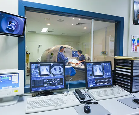
Technological basis of the MBST magnetic resonance therapy is MRI – magnetic resonance imaging
To put it simply, a magnetic resonance tomograph is a very large digital camera which takes photos of the inside of the body. The images show the different tissue structures in the body in such detail that medical professionals can see whether there is, for example, an inflammation in a "photographed" area or whether the tissue is healthy or ill, as in the case of tumours. To generate these images, hydrogen protons, a strong magnetic field, radio wave pulses and very powerful computers are needed. The patient is not exposed to any radiation during an MRI examination. But how does such an MRI work and why is the MBST magnetic resonance therapy system based on the same technological basis?
MRI technology explained simply

The magnetic resonance tomograph (also called magnetic resonance imaging or MRI for short) is usually a very large machine into which the patient is positioned. In its normally ring-shaped tunnel, often referred to as tube, a very strong magnetic field is generated to influence the hydrogen protons in the patient’s body. When the magnetic field is switched on, the hydrogen protons align themselves in a very specific way. The MRI device then sends out radio wave pulses that are precisely timed and stimulate the hydrogen protons by transmitting energy in such a way that their biophysical parameters are altered in a measurable way, i.e. the hydrogen protons are energetically pushed and change their position. When the pulses are switched off, the hydrogen protons return to their previous position. This can be measured from outside the body and the resulting data is transformed by a computer into pictures for diagnostic purposes.
But what does this have to do with MBST magnetic resonance technology and what exactly happens during therapeutic use?
Course of the diagnostic use of magnetic resonance technology
When a radio wave pulse is switched off, the hydrogen protons return to their energetic state of equilibrium, i.e. back to the position determined by the magnetic field. The time required for this process is called relaxation time. Part of the energy that the hydrogen protons have absorbed is then released and this is measured from outside the body. Since the water content of the different tissues in the body varies (content of water in bone tissue, for example, is lower than in cartilage), the tissues contain different amounts of hydrogen protons. Due to the different relaxation times of the different tissue types, MRI technology can represent these differences in the form of image contrasts. From the data of the measured signals and with the help of special mathematical methods, the computer generates an image in different shades of grey. By changing the measurement settings the presentation of certain types of tissue can be highlighted or softened, depending on the tissue that shall be examined.

Course of the therapeutic use of magnetic resonance technology
In MBST magnetic resonance therapy, a magnetic field is generated that prompts the hydrogen protons to align themselves according to the field lines. However, the generated field is many times weaker than that of an MRI. The reason is that MBST magnetic resonance technology only targets specific tissues and not all tissue structures in the target area. For example, MBST therapy sequences only stimulate hydrogen protons in tissues that are in resonance. These are the receivers of the energy transfer. In this way, various processes can be triggered and the metabolism of the cells can be influenced. Scientific data indicate that MBST magnetic resonance technology may stimulate various biophysical processes and trigger anti-inflammatory and pain-relieving effects.
Differences between the MBST magnetic resonance therapy system and magnetic resonance imaging (MRI)
The biggest difference is the use for diagnostic or for therapeutic purposes. In addition, the MBST magnetic resonance therapy system is characterized by the open design of the therapy devices. This means that patients do not have to fear suffering from claustrophobia during MBST treatment. They can relax and listen to music, read or even sleep.
The patented MBST magnetic resonance technology also requires no special premises and uses magnetic fields that are not nearly as strong as those of MRI. MedTec has succeeded in using the so-called Adiabatic Fast Passage to generate resonance conditions even at low magnetic field strengths. Therefore, contraindications for MRI do not necessarily apply to MBST therapy.
Why do MRT and MBST magnetic resonance technology use hydrogen protons?
Hydrogen is the most common element in the human body and at the same time the most sensitive component of the body for magnetic spin or magnetic resonance. By using hydrogen protons, the largest possible amount of protons in a tissue can be addressed.
History of the MRI
The theoretical foundations of magnetic resonance were laid in 1924 when the physicist Wolfgang Ernst Pauli described it for the first time. In 1946, Felix Bloch and Edward Purcell independently realized magnetic resonance, i.e. the resonance absorption of electromagnetic radiation by atomic nuclei located in a strong and high-frequency magnetic field, for which they were awarded the Nobel Prize in Physics in 1952.
In the 1970s, magnetic resonance imaging (MRI) was developed further based on the work of Peter Mansfield and Paul C. Lauterbur. It was not until 2003 that Paul C. Lauterbur and Peter Mansfield together received the Nobel Prize for Medicine and Physiology for their research into the imaging technology of magnetic resonance imaging (MRI), which has an immense diagnostic value. The imaging technology provides very important and accurate diagnostic images of body tissue while being gentle for the patient. Magnetic resonance imaging (MRI) is the gold standard of modern imaging diagnostics. In medicine, a diagnostic, therapeutic or general scientific procedure that represents the most proven and best solution in a given case is called gold standard. New methods are compared to that gold standard.
From MRI to MBST therapy

It was not until 1977 that R. Damian succeeded in creating the first image of the human body. The resolution was not yet sufficient for diagnostic use and the recording times were still several hours. In 1981, for the first time tumour tissue could be distinguished from healthy tissue. MRI was increasingly accepted clinically, among other things because of its advantages, e.g. the high soft tissue contrast and the lack of radiation exposure. In the early stages of MRI, patients often had to be examined many times. After frequent magnetic resonance imaging examinations, many Patients with joint problems reported – initially inexplicable to physicians – that their complaints had improved.
Axel Muntermann, the developer of therapeutic magnetic resonance, also became aware of these results. Together with physicians, biologists and physicists, he finally came to the conclusion that it might be the phenomenon of magnetic resonance that triggered this positive effect. Based on this, the MBST treatment systems, which use the same physcial principle as the MRI device – magnetic resonance – were developed in several years of work.
Advantages of MRI and MBST compared to CT, ultrasound or x-rays
MRI is one of the most modern, safe and gentle methods to detect pathological changes inside the body without the use of the burden of x-rays. With modern MRI scanners, location, extent and cause of a particular disease can be displayed much better than with conventional procedures such as x-ray examinations or ultrasound.
