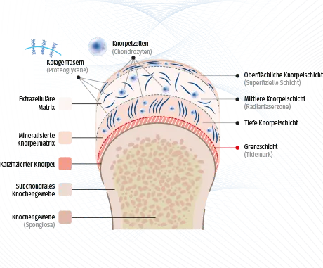
Cartilage cells · Chondrocytes
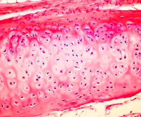
Cartilage consists mainly of specialized cells, the chondrocytes, and the extracellular matrix (ECM) produced by them. The extracellular matrix surrounds the chondrocytes. The proportion of chondrocytes in the total cartilage mass is only about one to ten percent. Their task is to maintain the homeostasis of the cartilage. Their shape is oval to round and they are located together in small groups. These cell groups and the extracellular matrix immediately surrounding them are known as chondrons.
Cartilage tissue is usually the weakest and at the same time most sensitive link in the human locomotor system. It is easy to damage, relatively susceptible to disorders and has no significant possibilities for repairing defects.
Since cartilage has no innervation, neither pain nor fatigue nor reaching the limits of resilience are perceived. However, cartilage also has no blood supply, which is why its metabolism must take place via diffusion, which is time-consuming. In addition, the cartilage tissue is relatively poor in cells, i.e. there are only a few chondrocytes. Consequently, they have to perform well in the production of extracellular substances. If individual chondrocytes do not fulfill this task optimally, the demands on the remaining chondrocytes increase.
Hyaline cartilage
Hyaline cartilage, with the exception of joint cartilage, is surrounded by a cartilage skin, the perichondrium. The hyaline cartilage in the joint has no perichondrium. During adolescence, its nutrition is accomplished through subchondrale vessels located in the bone. After maturation, however, calcification happens in the cartilage tissue and supply of the cartilage becomes possible only through the synovial fluid in the joint space. Therefore, cartilage tissue has only a small healing tendency.
The degradation of cartilage tissue caused by trauma or chronic and progressive changes is therefore a great challenge for doctors and researchers, since the poor regenerative capacity of human joint cartilage leads to osteoarthritis.
Cartilage cells can be chondroblasts, chondrocytes and chondroclasts. The precursor cells of chondrocytes, the chondroblasts, are called cartilage formers. These can synthesize all cartilage matrix components and therefore represent active cartilage cells. They are formed from mesenchymal stem cells. At the time when they stop this synthesis function, they become chondrocytes, the actual cartilage cells. These are smaller than the chondroblasts and contain a lot of water. The number, location and density of chondrocytes is specific for each type of cartilage.
Task
Function of the cartilage is to ensure the smoothest possible mobility of the joint surfaces and together with the joint fluid to provide pressure elasticity through optimum power transmission .
Cartilage degeneration
Nowadays, it is assumed that chondrocytes in osteoarthrotic cartilage begin to divide due to the loss of their matrix and under the influence of growth-regulating cytokines in order to maintain cartilage function. The augmented and increased division leads to the formation of so-called clusters (brood-capsules or growth-nests). These clusters then undergo destruction of the cartilage matrix, which is visible in deep fissures in the cartilage. A consequence can be necrosis of the cartilage cells and finally a complete abrasion down to the bone.
